
Oral Medicine & Radiology
Spotlight
Scope
- To give clinical diagnosis of oral diseases.
- To give clinical diagnosis of systemic diseases based on oral findings.
- Performing investigations and treatment planning in management of oral diseases.
- Radiological and imageological workup of oral diseases and give a radiological diagnosis.
- Non-surgical management of oral diseases where applicable.
- Dental management of medically compromised patients.
- Management of medical emergencies in dentistry.
NEW FRONTIERS:
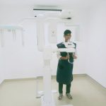
Advanced Imaging Centre
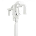
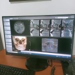
Cone-beam computed tomography (CBCT) is popularly known as “Third Eye in Dentistry”.It is an advanced radiological diagnostic tool for oral and maxillofacial region. It is a 3D Imaging technique used for diagnosis and treatment planning of various disorders affecting the Head and Neck Region of the body.
Manav Rachna Family believes in providing the best treatments at affordable prices with the state of art infrastructure, and with the addition of the CBCT is like a Feather in the Cap.
The Department of Oral Medicine and Radiology at,Manav Rachna Dental College, FDS,MRIIRSis now well equipped with a 3D Imaging CBCT Machine (Genoray 3D Papaya Plus, Unicorn).
INSTRUCTIONS FOR THE PATIENTS:
Cone beam CT examination requires no special preparation. Prior to the examination, the patient may be asked to remove metal objects, such as jewellery, eye-glasses, hairpins and hearing aids. Although removable dental work may need to be removed, it is advisable to bring these along, as the dentist may need to examine them. Women should always inform their dentist if there is any possibility that they are pregnant.
INDICATIONS OF CBCT:
IMPLANTS:
CBCT can give a clear picture about the quality of bone in which the implant is to be placed, the placement in relation to the surrounding structures and in cases where bone reconstruction may be required.

SURGICAL TREATMENTS:
This imaging modality allows for better diagnosis of tumors or cysts, the relationship of teeth to structures such as nerves, the presence and extent of impaction of teeth, extra teeth, foreign bodies, etc. With the correct knowledge about the presence, extent and relation of these structures

TRAUMA CASES:
CBCT is able to show clear fracture lines and fractures when compared to conventional images, depicting precisely the position and orientation of displaced fragments in a reasonably short time interval.
These could be tooth and socket fractures, fractures of the orbit, fractures of the jaws or any other structure of the face and oral cavity.

ORTHODONTICS:
CBCT is used for planning orthognathic surgeries, to evaluate the amount of airspace, to evaluate clefts in the palate, for analysis of facial structures, teeth and jaws, to place braces, to assess facial growth, age or disturbances in the eruption of teeth.

ENDODONTICS:
To evaluate the presence of superimposed or extra root canals, the presence of infection or pathologies around the tooth root, calcified canals, missed canals or extra canals.

PERIODONTICS:
CBCT allows for highly accurate analysis of bone loss as well as bone healing after periodontal treatment or regenerative therapy.

OUR TEAM
Our team of Oral and Maxillofacial Radiologists includes:
Dr. Sumit Bhateja, Dr. Tamanna Soni, Dr. Sheena Thamman
Orion made fully functional(Paperless functioning)
Dept. of Oral Medicine and Radiology, Manav Rachna Dental College has been monitoring Smooth functioning of Orion software for recording patients records at MRDC. This has greatly helped in the framework of MRDC’s functio
Digitization of Medical Records (Filmless Radiology)
Using VISTA Scan, a PSP based digital radiology technology has been implemented and integrated with Orion so that all radiographs are uploaded and attached to respective patient records. These can be viewed and downloaded by any department with access to the patient records.
By using a scanner at the OPD registration physical records are being recorded digitally in relevant cases.
Set up of Tobacco cessation center
Dept. of Oral Medicine and Radiology, Manav Rachna Dental College has established a Tobacco cessation center with a primary aim of providing timely intervention in cessation of tobacco related habits thereby reducing risk of many major diseases.
The center was started by Dr. Sumit Bhateja under the leadership of Dr. Arundeep Singh, Principal MRDC from 1st Oct. 2018 and till date more than 80 patients have been counseled & motivated to adapt to a Tobacco free lifestyle.
It comprises of Health Education room, counselling room & Tobacco clinic
CDE
The CDE programmes in the specialty focus on a multi disciplinary approach and encourage the participation from all departments. A CDE programme cum workshop titled “RESEARCH METHODOLOGY AND FUNDAMENTALS OF BIOSTATISTICS”was organized by the department on February 12th and 13th, 2015 with Dr. L. Nagesh, Dr. Giridhar J Gyani, Dr. R.M. Pandey and Dr. Arpit Gupta as guest speakers.
Infrastructure and Facilities
- A 100 kVp Extraoral machine has been added to the armamentarium for imaging of skull, chest and long bones.
- The department of Oral Medicine and Radiology at MRDC is equipped with the latest intra oral and extra oral (panoramic and cepahlometric) digital imaging systems in addition to 17 state of the art electronic dental chair/units. The radiology section has stringent radiation protection norms in place.
- The departmental library has adequate quality reading material in the form of books and journals to stimulate the inquisitive mind. These academic sources are used by the students and Interns to make regular scientific presentations in the department and is source of valuable information for the postgraduate students.
Our Courses

")


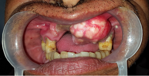
Case of the Month